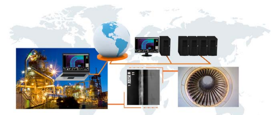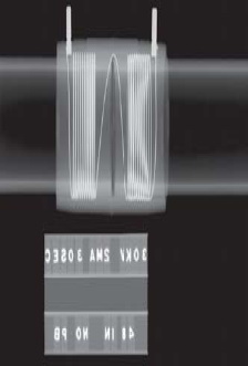
One of the most common and traditional methods of NDT is radiography. It is based on the registration of X-ray film ionizing radiation after its interaction with the object of control and analysis of the resulting image.
Despite the fact that radiography has long been used in the industry (the first X-ray laboratory, designed exclusively for industrial research, was established in 1925), so far it is an integral part of the production process.
Main advantages of the radiographic examination:
- high sensitivity to identify defects (average 1-2% of the rayed thickness);
- documentation of inspection results (radiographic film can be stored for many years);
- displaying the testing results (the image of the defect on the film can easily determine type of defect);
- applicability to a wide range of materials (depending on the source of ionizing radiation can be controlled, both metals, including austenitic steel and light metals, and organic matter).
Radiographic inspection modes of particular object depend on the sensitivity to radiation, contrast sensitivity and resolution of the converter used radiation (X-ray film), the intensity of the radiation source, and the geometric parameters of radiographic schemes. These modes have to be optimal sensitivity and productivity of monitoring. Energy of radiation, X-ray tube voltage should be chosen depending on the thickness and density of the inspected material.
To perform all the tasks described above, our company has a fleet of X-ray machines, both the permanent radiation and pulsed. Having such a large number of devices with different specifications allows to monitor objects made both of metallic and nonmetallic materials.

Depending on the object of control, control schemes and requirements an appropriate radiation source is selected for him to obtain the necessary results.
Formation of X-ray image on the film subject to the laws of geometrical optics, i.e., is fully analogous to the formation of shadows in visible light. Thus, the sharpness of an object image on the film depends directly on the size of the radiation source and the distance from it to the film and of the film to the object. Therefore, to obtain the most clear images, the film cartridge as close as possible to the controlled object. Controlled object and the film exhibited within a certain period, after which the film is removed and subjected to photographic processing. Photofinishing includes the steps of developing, fixing, washing and drying.
Radiographic film shows the best contrast and spatial resolution. However, along with the advantages of film radiography has number significant disadvantages:
- Low quantum efficiency;
- The narrow dynamic range;
- The long duration and complexity of the processing of the film material;
- Difficulties associated with the organization and content of the film archive.
To solve the above disadvantages of film radiography, our company has a digital complex Carestream Industrex HPX-1.
The method is based on obtaining an X-ray image on a plate coated with a special material to the phosphor. Flexible plate as x-ray film, set for object controlled. During exposure it accumulates energy ionizing radiation, thereby forming a latent image that can be stored for a long time (up to six hours). After completion of the exposure plate is placed in a scanner that reads the invisible image therein by a laser. Grayscale image is reproduced on the screen and directly perceived by the operator. The resulting digital image is optimized if necessary, scaled and stored.
Advantages of digital radiography compared with the film method.
- High-speed imaging;
- Elimination of time-consuming process in photochemicals film processing;
- Reduced doses required for exposure, as compared with the film;
- Wide dynamic range enables to explore material objects having a greater thickness or complex shape;
- Plate for imaging is reusable; permissible consistent exposure to tens of thousands of images;
- There is the ability to archive information on a variety of media, with virtually unlimited shelf life; and if you want to receive the required number of copies and use the network to transmit images.
Plates allow to directly receive digital images, bypassing the step of using the equipment to digitize X-ray films.
Significant disadvantages of the radiographic examination should be its X-ray radiation, which is ionizing, which has an effect on living organisms, and can cause radiation sickness and cancer. For this reason, when working with X-ray machines must observe the security measures.
In addition, the shortcomings of the radiographic examination should include the fact that the control cannot be detected discontinuities and inclusion:
- the size in the direction of less than twice the sensitivity of radiographic inspection;
- if their images in the images coincide with the images of foreign parts, sharp edges or sharp changes in thickness of the inspected metal.
